When dealing with spina bifida ultrasound detection, the use of prenatal imaging to identify spinal neural tube defects early in pregnancy. Also known as prenatal spina bifida screening, it relies on high‑resolution prenatal ultrasound, a non‑invasive sound‑wave exam that visualizes the fetus and often follows up with fetal MRI, magnetic resonance imaging that offers detailed soft‑tissue views. The detection process is closely tied to maternal folic acid, a vitamin supplement that dramatically reduces the risk of neural tube defects, and to the broader category of neural tube defects, congenital malformations of the brain and spinal cord. Understanding these entities helps you know what to expect from your screening journey.
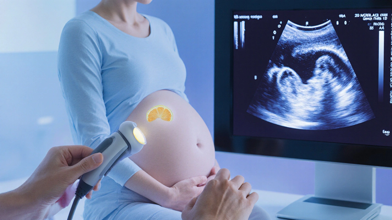
Spina bifida ultrasound detection encompasses three key steps: an initial anatomy scan around 18‑20 weeks, a targeted scan if the first exam shows suspicious features, and a confirmatory fetal MRI when needed. The anatomy scan uses a transabdominal probe to capture the “lemon sign” on the head and the “banana sign” on the cerebellum—classic markers that signal a possible defect. If these signs appear, a specialist sonographer may perform a detailed neurosonography, looking for the spinal defect’s level and severity. The next semantic triple: prenatal ultrasound requires a trained sonographer, because image quality hinges on skill, equipment, and gestational age. When the ultrasound suggests a severe open defect, clinicians often order a fetal MRI, which shines in distinguishing soft‑tissue details and assessing associated brain anomalies. Finally, the detection pathway links maternal folic acid supplementation to reduced spina bifida risk, so doctors will review your prenatal vitamin history and may recommend higher‑dose folic acid if you’re planning another pregnancy.
Beyond imaging, many providers combine ultrasound findings with maternal serum alpha‑fetoprotein (AFP) testing. Elevated AFP levels can hint at an open neural tube defect, prompting earlier ultrasound evaluation. This multimodal approach—ultrasound, AFP, and MRI—creates a robust diagnostic net, allowing families to make informed decisions about pregnancy management, surgical options, and post‑natal care. Early detection also opens the door to fetal surgery in select centers, where repairing the defect before birth improves neurological outcomes. As you navigate this process, remember that each test builds on the previous one, forming a clear picture of your baby’s health.
Below you’ll find a curated list of articles that dive deeper into each aspect—ultrasound technique, MRI interpretation, folic acid guidelines, and the latest in fetal surgery—so you can explore the details that matter most to you.
Learn how ultrasound spots spina bifida in unborn babies, the key sonographic signs, when fetal MRI adds value, and what preventive steps can lower risk.